
Luke Milliron, UCCE Orchards Advisor, Butte, Tehama & Glenn Counties; Dani Lightle, UCCE Orchards Advisor, Glenn, Butte & Tehama Counties; and Franz Niederholzer, UCCE Orchards Advisor, Sutter-Yuba & Colusa Counties
In prune production, a new mantra has become “some years are bacterial canker years, while every year is a Cytospora year”. Bacterial canker infections are caused by Psyudomonas syringae (same bacterium that causes bacterial blast in almond). While P. syringae is ubiquitous across surfaces in the orchard, it only causes infections and damage in certain years, under the right environmental conditions. Wet/cold springs are conducive to bacterial canker infections, which although severe and often lethal, do not continue to spread the following year (i.e. annual disease, not perennial). Going into this spring, we knew the wet/cold conditions in late winter could mean that we were likely headed into a bacterial canker year. However, diagnosing the dieback (cankers) we are seeing this spring has brought some surprises.
The branch dieback we are seeing in prune orchards have key similarities and differences we used to help diagnose the source of the issue.
CASE 1. Two adjacent prune blocks: one unaffected, and one affected block that has aggressive dieback (photos 1-2). Affected branches show the classic mottled cankers diagnostic of bacterial canker (photo 3). A key difference between the blocks is that the unaffected block was treated for nematodes. Ring nematode is a predisposing-factor to bacterial canker. Samples from this orchard were analyzed for fungal pathogens and results included the silver leaf pathogen (Schizophyllum commune), Fusarium, and Botryosphaeria. Since bacteria were not included in this analysis, the original cause of the dieback could still be oil burn from a dormant application and bacterial canker caused by P. syringae.
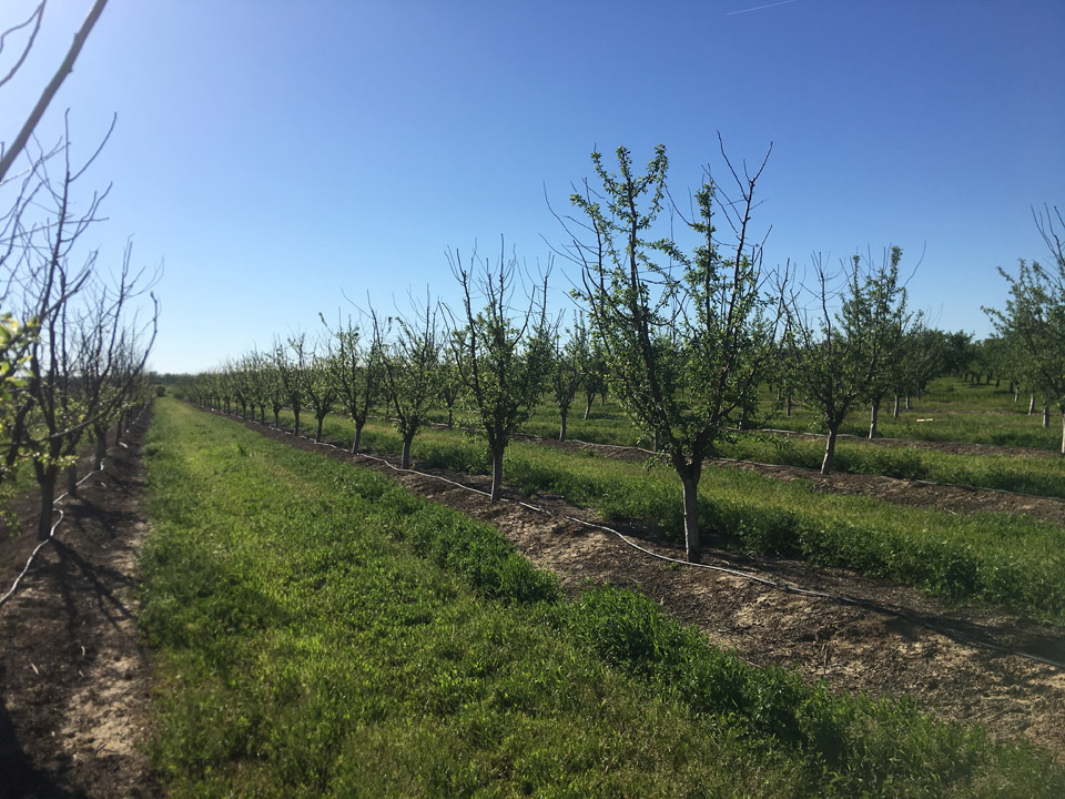
Photo 1. The block on the left had extensive limb dieback this spring. One of the differences in management between the two blocks is that the one on the right was treated for nematodes, ring nematode is a predisposing stressor for bacterial canker infections (photos: Franz Niederholzer).
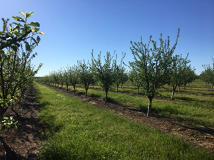
Photo 2. This block on the right did not suffer dieback. One of the differences in management between the two blocks is that the one on the right was treated for nematodes, ring nematode is a predisposing stressor for bacterial canker infections (photos: Franz Niederholzer).
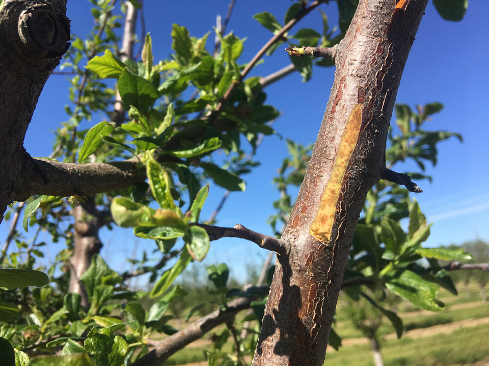
Photo 3. Classic mottled damage often associated with bacterial canker infections (photo: Franz Niederholzer).
CASE 2. Many trees in the orchard show discrete dieback of upright branch tips that is consistent with how oil burn presents (photos 4-6). Oil was applied to this block during dormancy (2-3 gallons December/January). Although oil can help provide effective control of scale insects, drying weather after application (e.g. a drying north wind) can lead to phytotoxicity “burn” and dieback. One edge tree in the orchard also showed the classic bacterial canker symptoms of extensive dieback with rough bark cankers (photo 7). Being an edge tree, it could have experienced a higher rate of applied oil. However, as an edge tree it is also subject to countless other potential stressors that could predispose the tree to bacterial canker.
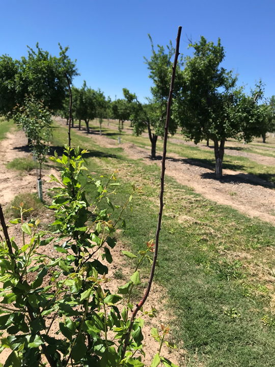
Photo 4. An orchard with branch tip dieback suspected to be from a dormant oil application. (Photo: Luke Milliron).
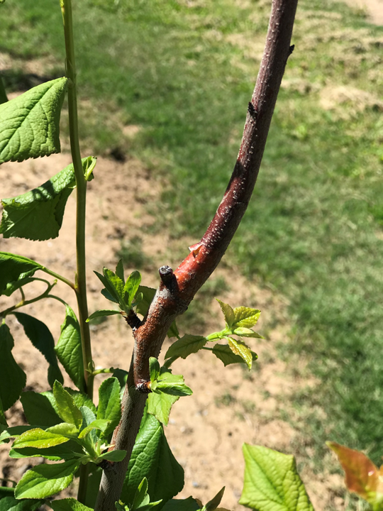
Photo 5. Regrowth below the dieback in an orchard with branch tip dieback suspected to be from a dormant oil application. (photo: Luke Milliron).
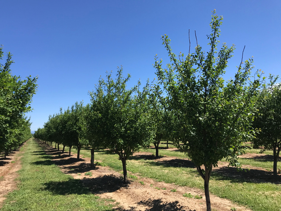
Photo 6. Orchard with branch tip dieback suspected to be from a dormant oil application (photo: Luke Milliron).
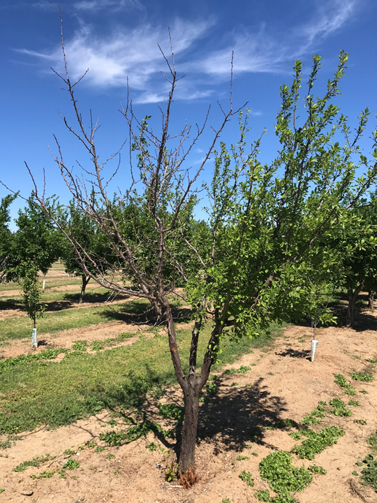
Photo 7. One edge tree in the orchard also showed the classic bacterial canker symptoms of extensive dieback with rough bark cankers (photo: Luke Milliron).
CASE 3. Select patches of trees in this orchard show the extensive dieback consistent with bacterial canker (photo 8). Oil was also applied during dormancy in this orchard (2-3 gallons December/January). Bacterial canker was very likely the cause of this dieback due to the tell-tale signs of flecks (photo 9) and discrete patches (photo 10) of canker, as well as the fermented/sour-smell associated with the sour sap phase of bacterial canker decline.
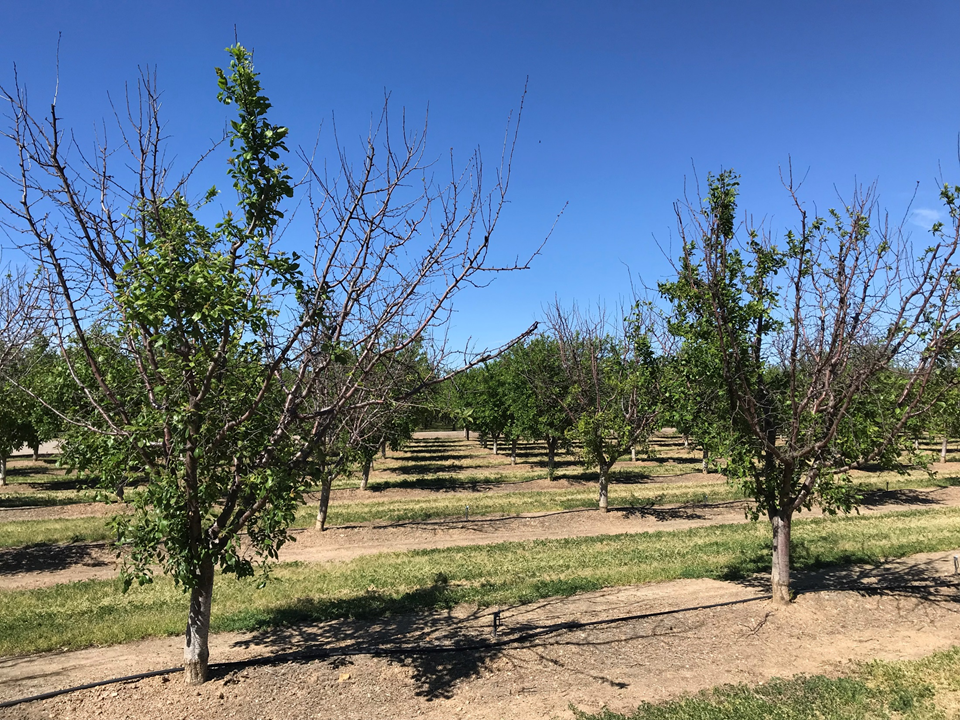
Photo 8. Select patches of trees in this orchard show the extensive dieback consistent with bacterial canker (photo: Luke Milliron).
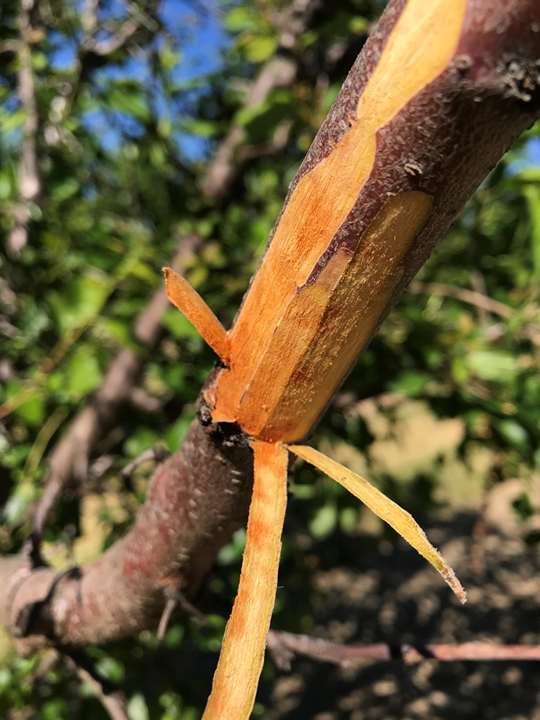
Photo 9. Bacterial canker was very likely the cause of this dieback due to the tell-tale signs of flecks, as well as the fermented/sour-smell associated with the sour sap phase of bacterial canker decline (photos: Luke Milliron).
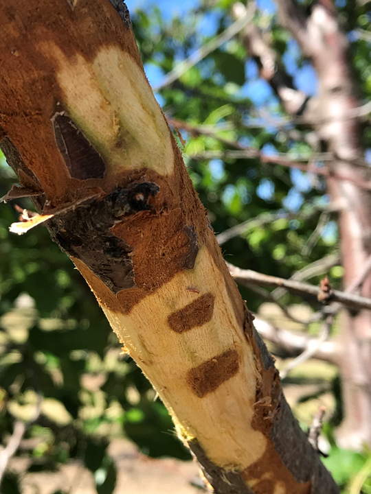
Photo 10. Bacterial canker was very likely the cause of this dieback due to the tell-tale signs discrete patches of canker, as well as the fermented/sour-smell associated with the sour sap phase of bacterial canker decline (photos: Luke Milliron).
CASE 4. Orchard with canker dieback was first noticed last year when trees were not flowering out, except low in the canopy. These symptoms were most commonly in trees planted on vigorous rootstocks. The vigorous rootstocks (e.g. Atlas, photos 11-13) would have had the largest cuts made to it when mechanical (boxed) pruning cuts were made in the fall of 2017. These pruning cut in the fall of 2017 are presumed to be the source of Cytospora infections during subsequent rainfall events. Approximately 30 days of dry conditions are required after a pruning cut is made for the surface to callus over and not be susceptible to infection. Topsin-M®, has been shown to have efficacy in protecting pruning wounds and reducing Cytospora canker incidence in prunes. Dr. Themis Michailides (UCCE Kearny Ag Center) identified Cytospora from branch samples in May 2018.
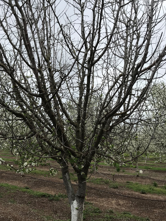
Photo 11. Orchard with canker dieback was first noticed last year when trees were not flowering out, except low in the canopy. Photo: Luke Milliron.
During the 2019 bloom, extensive dieback was identified on some trees (photo 11) and one tree was cut back to see how extensive the canker damage had spread. The canker damage continued to grow down to a primary scaffold (photo 12). The cut branches showed the classic bark canker margin of Cytospora and other bark cankers (see red arrow in photo 14). There was also internal dead tissue that was not spreading along the bark (arrow photo 15); this was presumed to be Phellinus or another heart rot species. However, the diseased wood came back with 100% Cytospora. This was the first time that Michailides had seen Cytospora as a canker internal to the bark.
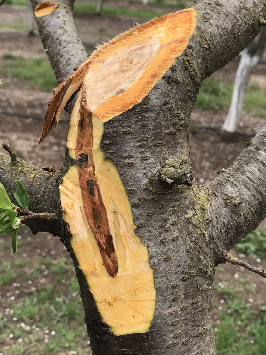
Photo 12. Cutting back one tree to see how extensive the canker damage had spread showed that damage continued to grow down to a primary scaffold. Photo: Luke Milliron.
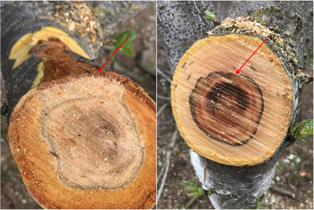
Photo 13. Cut branches showed the classic bark canker margin of Cytospora and other bark cankers (see red arrow, left photo). There was also internal dead tissue that was not spreading along the bark (red arrow, right photo); this was presumed to be Phellinus (i.e. heart rot). However, the diseased wood came back with 100% Cytospora. This was the first time that Michailides had seen Cytospora as a canker internal to the bark (photos: Luke Milliron).
CASE 5 (photo 14). Wood dieback, generally on smaller wood high in the canopy on some select trees in this grower’s field was sampled and sent into the Michailides Lab in mid-April. The analysis came back as one branch with phytotoxicity and the others with varying percentages of Botryosphaeria/Cytospora/Paecilomyces/Phomopsis. These results might suggest secondary fungal invasions following damage from an oil application; however the grower did not apply any oil. The grower also generally has an excellent program for potassium (avoiding potassium burn dieback), as well as for aphids and other potential causes of sunburn and subsequent fungal canker invasion. The grower believes the trees with dieback are on sandier parts of the orchard, but few other clues are helping us to figure this case out.
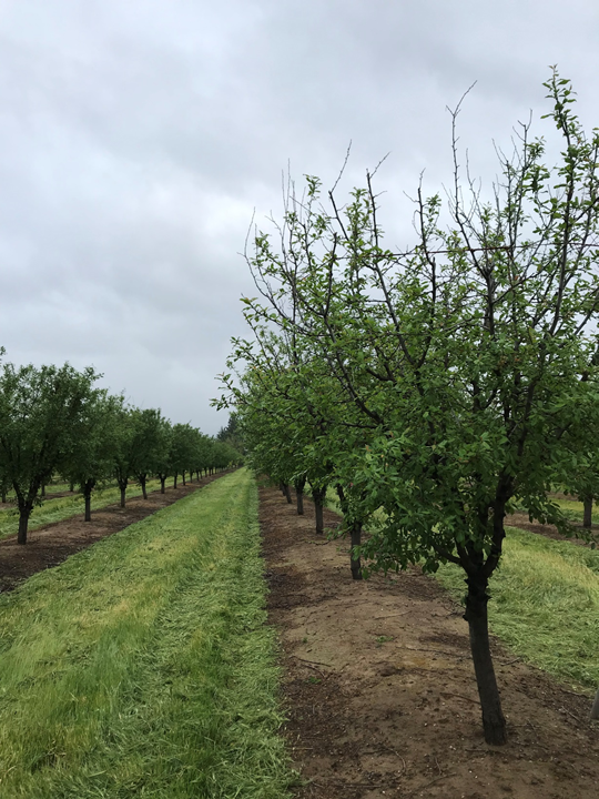
Photo 14. Wood dieback, generally on smaller wood high in the canopy on some select trees in this grower’s field. Several fungal canker diseases were isolated from sampled branches, and the underlying cause of the infections remains unknown (photo: Luke Milliron).
These six cases depict the expected prevalence bacterial canker incidence following a wet/cold spring. These cases also revealed that certain patterns of branch dieback are much easier to diagnose than others! We want to thank Dr. Michailides Lab for their support in diagnosis.
Leave a Reply